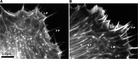
Figure 1.
Examples of actin cytoskeleton phenotypes in fish fibroblasts showing differences in the lamella region (LM) behind the anterior zone containing lamellipodia (LP) and filopodia (FP). Cells were transfected with mCherry-actin. Images show single frames from video sequences. (A) Lamella with bundles parallel to the cell front, together with arc shaped segments (AC). (B) Lamella with stress fiber bundles (SF) mainly perpendicular to the cell front. See Videos 1 and 2 (available at http://www.jcb.org/cgi/content/full/jcb.200709134/DC1). Bar, 10 μm.

