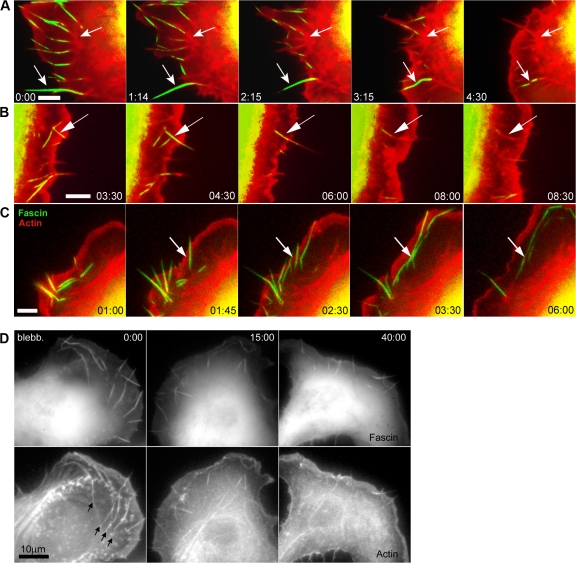
Figure 9.
Contribution of microspikes in B16 melanoma cells to construction of actin bundles in the lamella network. (A–C) Video sequences of cells expressing GFP-fascin and mCherry-actin showing transition of fascin-positive microspikes (arrows) into radial (A and B) and transverse bundles (C, arcs) in the lamella. (D) Inhibition of myosin contractility by 30 μM blebbistatin in a B16 melanoma cell expressing GFP-fascin and mCherry-actin (see Video 10). The fascin images were obtained only at the given time points shown in the video sequence to avoid inactivation of blebbistastin. The arc-shaped bundles arising from the lateral translation of microspikes (arrows at time 0:00) dispersed in blebbistatin, but microspike segments continued to enter the lamella (times 15:00 and 40:00). Times are given in minutes and seconds.

