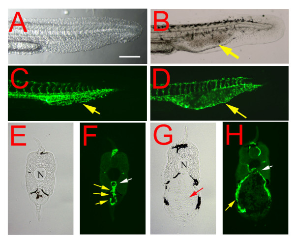
Figure 3.

Embryonic phenotype of the reg6 mutation. (A, C) In a 36-hpf (hours post fertilization) wild-type embryo (A, bright field; C, green fluorescent), the developing caudal veins form vascular plexuses (yellow arrow in C). (B, D) In contrast, endothelial cells of reg6 embryos (B, bright field; D, green fluorescent) develop fewer vessel branches and form swollen lumina (yellow arrows). Lateral view; anterior, left; dorsal, top. Scale bar, 100 μm for A-D. (E-H) Cross-sections of the tail regions showing multiple lumina of the capillary plexus of a wild-type caudal vein (E, yellow arrows in F) but a single enlarged lumen in reg6 (G, yellow arrow in H). N, notochord; white arrows, dorsal aorta; yellow arrows, caudal vein; red arrow, accumulated blood cells.
