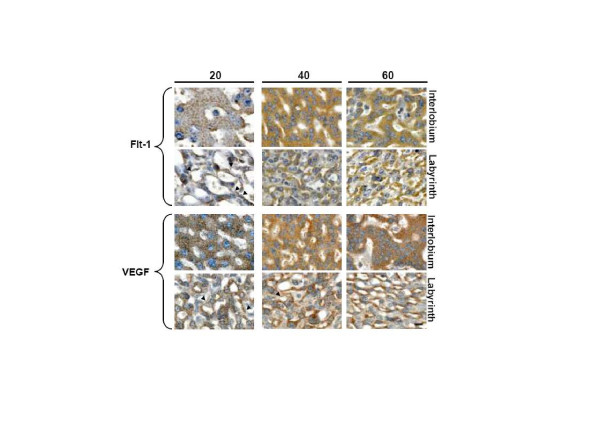
Figure 3.
Syncytiotrophoblasts composing the interlobium and the labyrinth expressed Flt-1 and VEGF, in sections obtained in days 20, 40 and 60 of pregnancy at a high magnification (1000×). The immunoreactivity showed a granular pattern for interlobar Flt-1, and for interlobar and labyrinthine VEGF; labyrinthine Flt-1 displayed a diffuse cytoplasmic staining. Arrowheads highlight linear signal in endothelial cells. Bar = 100 μm.

