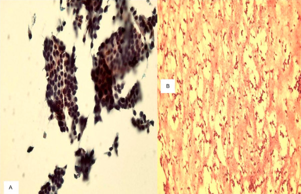
Figure 4.

a. A TP slide of follicular neoplasm on thyroid FNA showing cellular specimen and groups and 3-dimentional clusters of follicular cells with hyperchromatic nuclei. b. The cell block section of the same case shows acellular specimen (400×, Papanicolaou and H&E stains).
