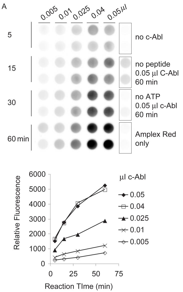
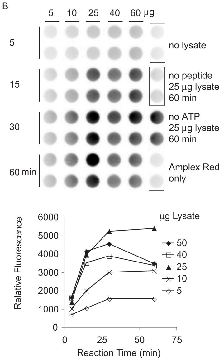
Fig. 3.
Dependence of kinase reaction on reaction time and kinase amount. Peptides (0.05 mM of Cys-AT and Cys-AL) were tethered to hydrogels in wells as describe in Materials and Methods. (A) Recombinant c-Abl: Kinase reactions were performed with 0.005, 0.01, 0.025, 0.04 and 0.05 μl c-Abl (0.1U/μl) in kinase buffer for 5, 15, 30, and 60 min. (B) K562 lysate: Kinase reactions were performed with 5, 10, 25, 40, or 50 μg of K562 cell lysate in kinase buffer for 5, 15, 30, or 60 min. Each panel shows a scan of Amplex Red reaction product on the top and a plot of total well fluorescence on the bottom. Control wells on the right are pairs of mock reactions lacking kinase (c-Abl or Bcr-Abl), lacking peptides, lacking ATP, or empty wells to which the Amplex Red detection reagent was added.


