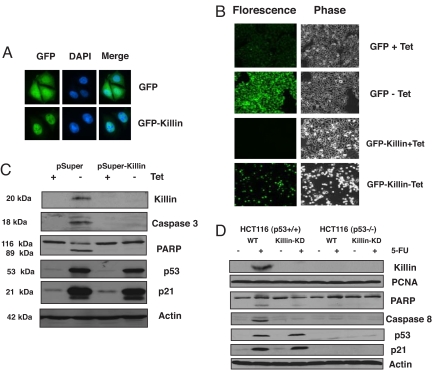
Fig. 3.
Killin is localized in the cell nucleus and is both necessary and sufficient for p53-mediated apoptosis (A) DLD-1 cell lines stably expressing either inducible GFP or GFP-Killin was visualized by fluorescence microscopy after 16 h of induction (−tet). Note that GFP-Killin is localized exclusively in the nucleus, whereas GFP is expressed throughout the cells. DAPI was used to stain cell nuclei. (B) Inducible GFP-Killin expression causes massive cell apoptosis and detachment within 72 h. Inducible GFP alone served as a negative control. (C) RNAi knockdown of killin expression blocks extrogenous p53-induced Caspase-3 activation and PARP cleavage. p53–3 cells stably transfected with either pSuper vector alone or pSuper-Killin were either noninduced (+tet) or induced (−tet) for p53 for 24 h. The induction of p53 and p21 was confirmed by Western blot analysis with β-actin as a control for equal sample loading. RNAi knockdown of killin expression led not only to diminished p53-dependent nuclear Killin protein expression but also to inhibition of Caspase-3 activation and cleavage of PARP as analyzed by Western blot. RNAi knockdown of killin expression had little effect on p53 induction and its effect on p21 expression. (D) RNAi knockdown of killin expression blocks endogenous p53-induced caspase-8 activation and PARP cleavage. HCT116 cells with or without endogenous p53 were infected with pSuperRetro-killin RNAi expression vector (Killin-KD). Cells stably selected for RNAi expression were treated or mock-treated with 5-FU (50 μg/ml) and 50 μg of either nuclear protein (for Killin and PCNA) or cytoplasmic protein extracts were analyzed by Western blot with corresponding antibodies, as indicated.

