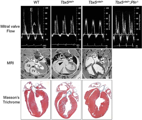
Fig. 1.
Diastolic dysfunction in Tbx5 ventricle-specific haploinsufficient mice. (Top) Doppler signal at the MV showing E and A waves. The E wave is decreased, and the A wave is increased in Tbx5del/+ and in Tbx5Vdel/+ mice. These values are restored to normal in Tbx5Vdel/+;PLN−/− mice. (Middle) MRI showing ASDs (arrowhead) in Tbx5del/+ but not in Tbx5Vdel/+ mice. la, left atrium; lv, left ventricle; ra, right atrium; rv, right ventricle. (Bottom) Histology of adult hearts stained by Masson's trichrome.

