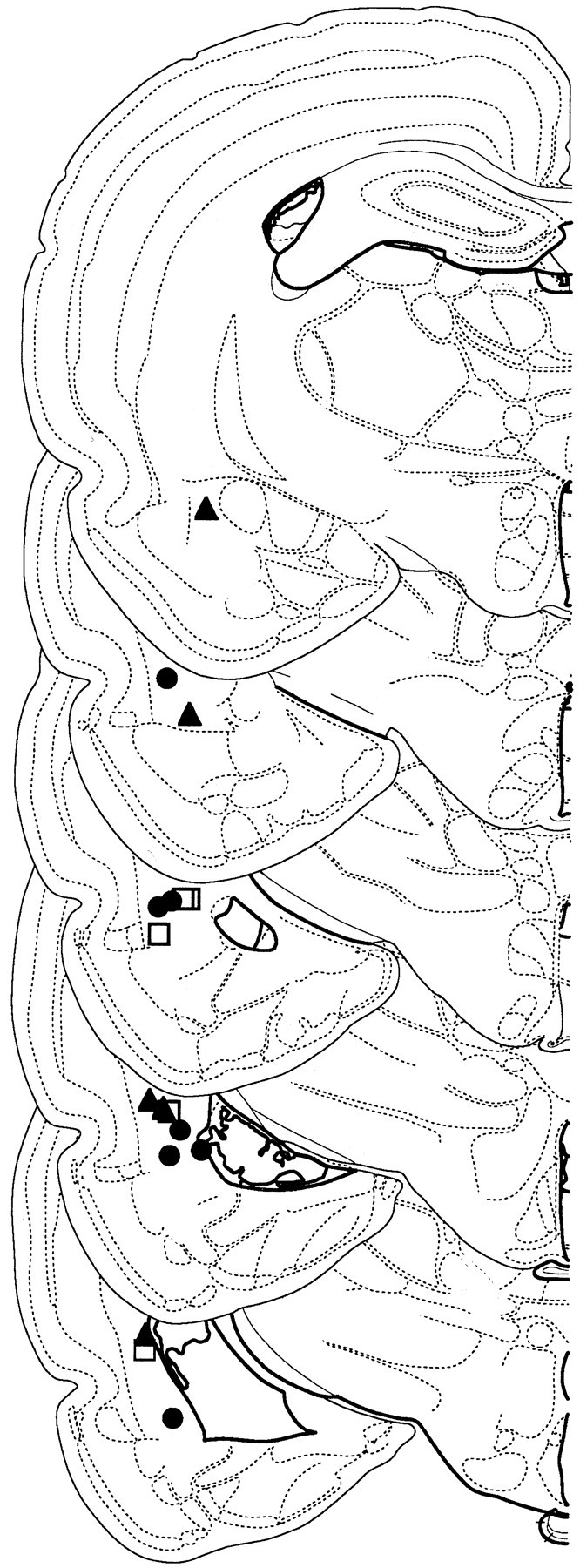
Figure 1.

Recording electrode placements in lateral amygdala. Placements represent all NO-EXT (filled triangles), HAB (open squares), and EXT (filled circles) rats included in the final analysis. Schematic brain section images are displayed from most rostral (top; -2.0 mm posterior to bregma) to most caudal (bottom; -3.9 mm posterior to bregma). Images were adapted from Swanson (1992).
