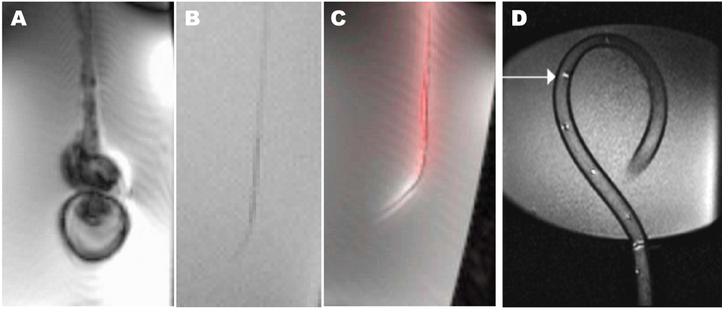
Figure 1.

Comparison of catheter visibility in vitro using steady-state free precession MRI. (A) A conventional steel-braided catheter disturbs the local magnetic field and distorts the image. (B) Removing the steel braid renders the catheter indistinct under MRI because it has no excitable spins to produce signal. (C) The same catheter, converted into an “active” antenna, detects excited surrounding spins and now is highly visible. Conspicuity is enhanced by post-processing techniques including gain adjustment and color highlighting. (D) A different active catheter device (Hegde et al, 2006) using multiple tuned microcoils that can be used in profiling mode or tracking mode.
