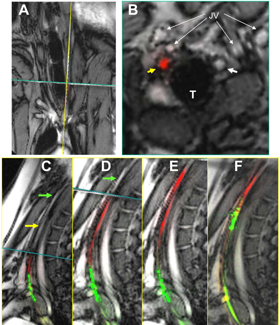
Figure 3.

Recanalization of chronic total occlusion using an active catheter and active guidewire. (A) Real-time, multiplanar MRI of the occlusion. (B) Transverse image shows the guidewire (red) traversing the occlusion (yellow arrow). (C–F) A sequence showing guidewire recanalization and catheter advancement. (C) The guidewire (red) engages the segment of interest, (D) traverses the occlusion, (E) and enters the patent distal carotid artery. (F) The catheter (green) is then advanced over the wire and across the occlusion. From Raval et al, 2006.
