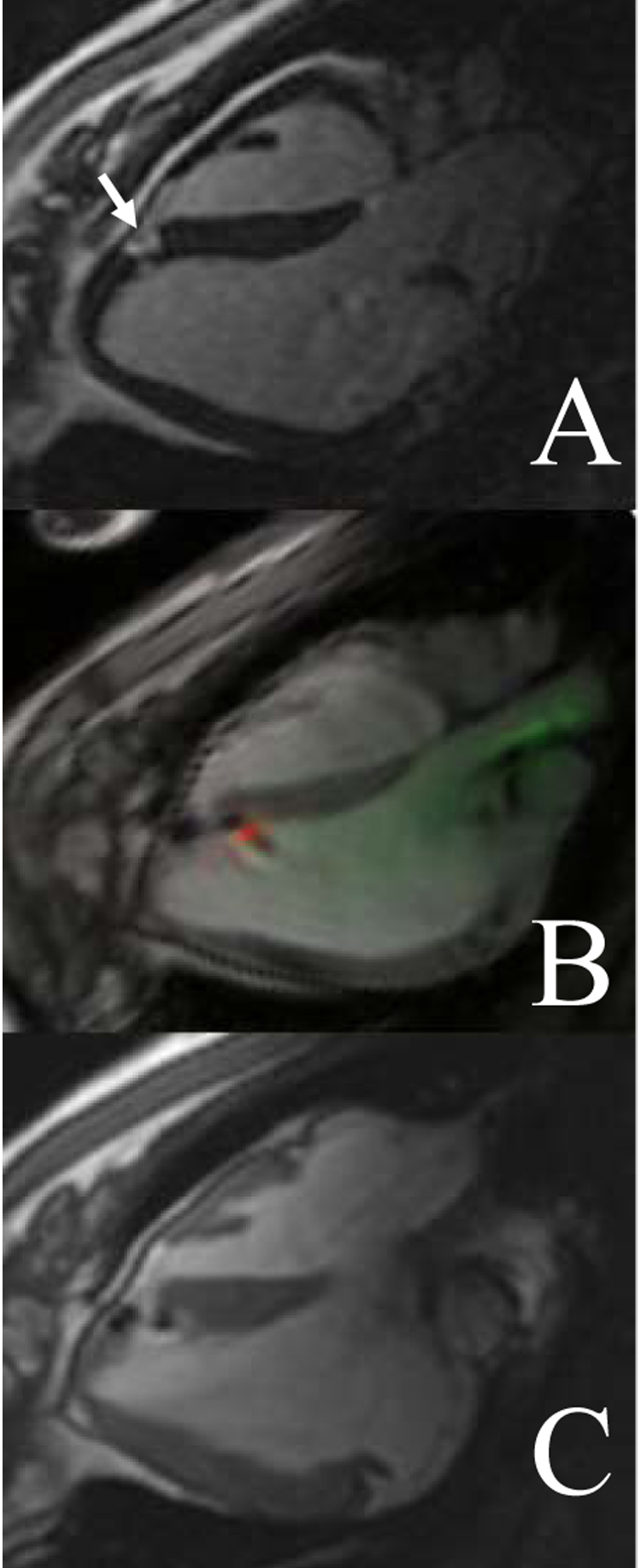
Figure 5.

Precise targeting of small infarct for MR-guided endomyocardial cell delivery. (A) Delayed gadolinium enhancement shows a small infarct (arrow) measuring approximately 1 cm2. (B) Real-time imaging shows an active endomyocardial injection catheter (green) and needle (red) delivering cells labeled with iron oxide. (C) The cells (dark spots) are delivered to the infarct borders. From Dick et al, 2003.
