Figure 5.
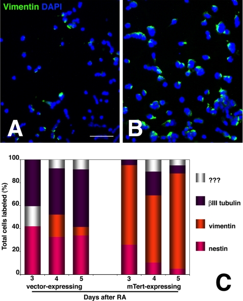
Persistence of vimentin immunoreactivity in differentiating mTERT-expressing lines. (A and B) Low power fluorescent photomicrographs of transfected P19 lines 3 d after RA treatment that have been reacted with anti-vimentin mAb. Nuclei were counterstained with DAPI. (A) Control line P2 (line P3 appeared similar); (B) mTERT-expressing line A5 (line A6 appeared similar). The mTERT-expressing lines show significantly more vimentin labeling. Bar 250 μm. (E) The percentage of total (DAPI-labeled) cells expressing vimentin in the control and mTERT expressing lines was averaged on days 3–5 after RA (orange) and then plotted in comparison with Nestin (pink), βIII-tubulin (purple), and non-RA responsive (white). Significantly, more cells from the mTERT-expressing lines labeled with vimentin, and those cells retained label past the time when neurons normally mature.

