Figure 9.
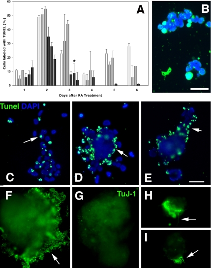
Fate of mTERT-expressing lines in aggregate cultures. (A) Quantitation of TUNEL labeling in aggregated cells from P19 parent cells (□) the control lines P2 and P3 (▩) and the mTERT-expressing lines A4, A5 and A2 (■). Both the parent P19s and the control lines undergo apoptotic events at days 2 and days 5 and 6, whereas apoptosis in the mTERT-expressing lines is reduced by 30%. (*p < 0.05 after ANOVA analysis followed by Student's t test compared with parent and P3 lines). (B) P2 cells treated with TUNEL to transfer biotin-labeled dUTP to cleaved DNA in apoptotic cells. Cell nuclei counterstained with DAPI. Bar, 50 μm. (C–I) Transfected ES cells 8 d after treatment with RA dissociated to complete differentiation. (C–E) Tunel labeling. Cell nuclei counterstained with DAPI. (C) Control line (E1), apoptotic cells are scattered throughout the aggregate (arrow). (D and E) mTERT-expressing lines M2 and M3. Apoptotic cells are concentrated at the aggregate perimeter. Bar, 500 μm. (F–H) TuJ-1 labeling to detect βIII-tubulin expression at 8 d after RA treatment. (F) ES control line E1. Cells at the perimeter of aggregates are heavily labeled with TuJ-1, indicating that they are differentiating neurons (arrow). (G–I) mTERT expressing lines M2 and M3. (G) No TuJ-1–labeled neurons migrating out from the perimeter of the M2 aggregate. (H and I) TuJ-1–labeled cells that remain within residual aggregates differentiate and send out neuritis (arrows). Bar, 500 μm.

