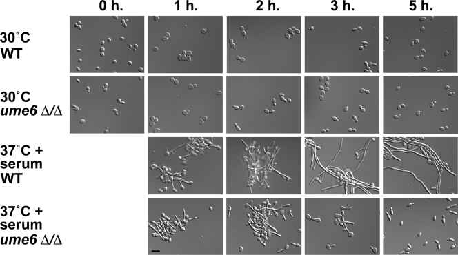
Figure 2.
Morphology of wild-type and ume6Δ/Δ cells undergoing the blastospore to filament transition. Wild-type (WT) and ume6Δ/Δ strains grown under non–filament-inducing conditions (YEPD medium at 30°C) were diluted into pre-warmed YEPD medium at 30°C or 37°C in the presence or absence of 10% fetal calf serum (FCS). Induction time (h) is shown on top. Aliquots of cells were fixed in 4.5% formaldehyde, washed twice in 1× PBS, and then visualized by Nomarski/DIC optics. Please note that the 0-h time point shows cells immediately before induction. Bar, 10 μm.

