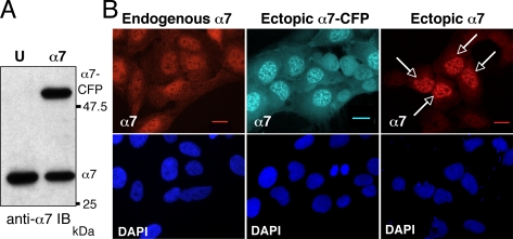
Figure 1.
Ectopic α7-CFP and α7 accumulate into nuclear foci. (A) Expression of the α7-CFP protein in cells. Total cellular extracts (30 μg) of human osteosarcoma U2OS-tTA (U) or U2OS-tTA cells stably transfected with vectors conditionally expressing α7-CFP (α7) cultured in the absence of tetracycline for 48 h were separated by electrophoresis and analyzed by immunoblotting using anti-α7 antibodies. (B) The cellular distribution of α7 in U2OS-tTA cells either untransfected (endogenous α7), stably transfected with pTRE2-α7-CFP (ectopic α7-CFP), or transiently transfected with pcDNA3-α7 (ectopic α7) was analyzed by indirect immunofluorescence using anti-α7 antibodies (red) or by CFP fluorescence (cyan) on fixed cells. Arrows indicate transiently transfected cells. Observation with a 63× objective. Bar, 10 μm.

