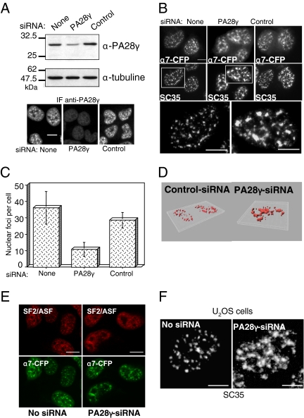
Figure 8.
Proteasome-PA28γ complexes are required for the integrity of the NS. (A) U2OS-tTA-α7-CFP cells were induced for α7-CFP expression and concomitantly transfected with control-siRNA or PA28γ-siRNA (siRNA 1) duplexes. Cells were recovered 48 h after transfection, and the expression level of PA28γ was analyzed either by immunoblotting or by indirect immunofluorescence using an anti-PA28γ antibody. Bar, 10 μm. (B) NS were visualized by the fluorescence of α7-CFP and by indirect immunofluorescence using anti-SC35 antibodies in induced U2OS-tTA-α7-CFP cells untransfected or transfected with PA28γ- or control-siRNA duplexes. Enlargements of the marked areas are presented. Bar, 10 μm. (C) Quantification of the number of α7-CFP–labeled NS in cells treated with PA28γ- or control-siRNA duplexes. Quantification was performed visually, by counting on the pictures the number of compact fluorescent foci in each cell. The values correspond to the means of five independent experiments (n = 100 cells, ±SD). (D) 3D reconstruction. SC35 localization was detected by indirect immunofluorescence in induced U2OS-tTA-α7-CFP cells treated with control- or PA28γ (1)-siRNA. Fixed cells were observed with a Leica DMRA microscope equipped with a 63× PL APO (NA = 1.32) oil immersion objective and a N2.1 (Leica) filter set. Stacks of images were acquired using a piezo stepper (Physik Instruments, Waldbronn, Germany), Metamorph 7.1 (Molecular Devices, Menlo Park, CA) and a Micromax 1300YHS CCD camera (Princeton Research Instruments, Princeton, NJ). Stacks were further deconvolved using a MLE algorithm and the Huygens 2.9 software (Scientific Volume Imaging, Hilversrum, The Netherlands) and analyzed in 3D using Imaris 5.3 (Bitplane, Zurich, Switzerland). (E) NS were visualized by the fluorescence of α7-CFP and by indirect immunofluorescence using anti-SF2/ASF antibodies in induced U2OS-tTA-α7-CFP cells untransfected or transfected with PA28γ- or control-siRNA duplexes. Bar, 10 μm. (F) In parental U2OS-tTA cells, untreated or treated with PA28γ-siRNA duplexes, NS were visualized by indirect immunofluorescence using anti-SC35 antibodies. Bar, 10 μm.

