Figure 4.
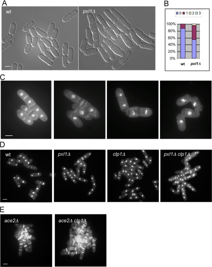
pxl1Δ cells have cell separation defects and display genetic interaction with clp1Δ. (A) Cell separation defects in pxl1Δ mutant. Wild-type and pxl1Δ mutant cells were grown in YES medium at 24°C and observed by light microscopy. (B) Quantification of the percentage of cells with septa as well as the number(s) of septa in wild-type and pxl1Δ mutant cells (n = 500). (C) Examples of abnormal cells in the pxl1Δ mutant. pxl1Δ cells were grown at 24°C, and samples were fixed and stained with aniline blue and DAPI to visualize septa and nuclei, respectively. (D) pxl1Δ clp1Δ double mutant cells accumulate multiple nuclei in the same compartment. Cells of indicated genotypes were grown in YES medium (or YES plus 1.2 M sorbitol medium) at 24°C. Samples were fixed and stained with aniline blue and DAPI to visualize septa and nuclei, respectively. (E) ace2Δ clp1Δ double mutant cells do not accumulate multiple nuclei in the same compartment. Cells of indicated genotypes were treated as D. Scale bar, 5 μm.

