Figure 5.
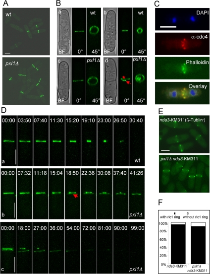
pxl1Δ mutant cells have a defect in the organization of the actomyosin ring. (A) pxl1Δ mutant cells have a double ring structure. Wild-type and pxl1Δ mutant cells expressing rlc1-GFP were grown at 24°C and observed by confocal microscopy. Z-stack images were taken and 3D reconstruction images with maximal projection are shown. (B) pxl1Δ mutant cells have a single unconstricted ring at early mitosis and a double ring at late stages. Individual cells from A were observed by confocal microscopy, Z-stack images were taken, and 3D reconstruction images are shown at different angles. Cells a and c, early stage. Cells b and d, late stage. Arrow indicates the primary ring and star indicates the secondary ring. (C) Cdc4p and F-actin are present in the double ring structure. pxl1Δ cells were grown in YES medium at 24°C. Samples were fixed and stained with DAPI, α-cdc4 antibodies, and phalloidin to visualize nuclei, Cdc4p, and F-actin, respectively. (D) Formation of the secondary ring structure in pxl1Δ mutant cells. Wild-type cells expressing rlc1-GFP were grown at 24°C and observed by time-lapse confocal microscopy. Z-stack images were taken at different time points, and 3D reconstruction images with maximal projection are shown panel a. pxl1Δ cells expressing rlc1-GFP were treated and observed as above (b and c). Arrow indicates the appearance of the secondary ring structure. (E) Formation of metaphase actomyosin ring is normal in pxl1Δ mutant cells. nda3-KM31 and nda3-KM311 pxl1Δ cells expressing rlc1-GFP were grown at 32°C to log-phase and shifted to 19°C for 6.5 h to arrest cells at metaphase. Images were taken by fluorescence microscopy. (F) Quantification of the number of metaphase actomyosin rings in wild-type and pxl1Δ mutant cells. Cells from E were quantified for the presence of actomyosin rings (n = 200). Time is indicated in min::sec. BF, bright field. Scale bar, 5 μm.

