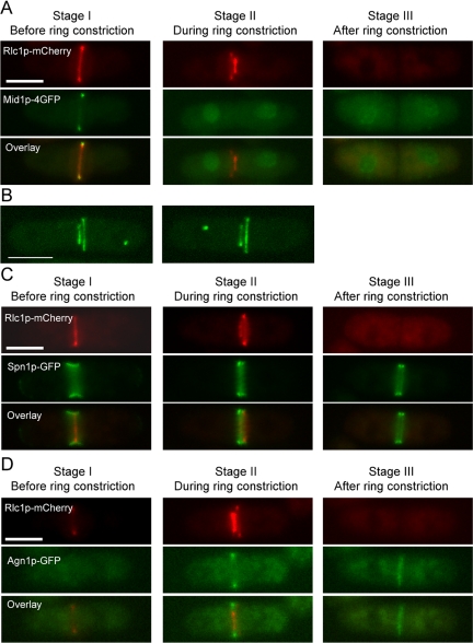
Figure 6.
Assessment of localization of various cytokinetic proteins to the secondary rings in pxl1Δ mutant cells. (A) Mid1p localizes to the nucleus during the primary ring constriction in pxl1Δ mutant cells. pxl1Δ cells expressing rlc1-mcherry mid1-4GFP were grown at 24°C to log-phase. Samples were taken and observed by fluorescence microscopy. (B) Cdc7p was distributed randomly with respect to the primary rings in pxl1Δ mutant cells. pxl1Δ cells expressing rlc1-GFP cdc7-GFP were grown at 24°C to log phase. Samples were taken and observed by confocal laser microscopy. Z-stack images were taken and 3D reconstruction images with maximal projection are shown. (C) Spn1p localizes to the primary ring in pxl1Δ mutant cells. pxl1Δ cells expressing rlc1-mcherry spn1-GFP were treated as A. (D) Agn1p localizes to the primary ring in pxl1Δ mutant cells. pxl1Δ cells expressing rlc1-mcherry agn1-GFP were treated as A. Scale bar, 5 μm.

