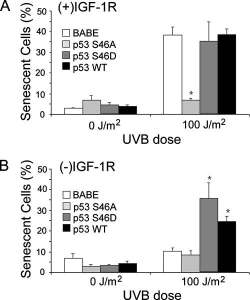
Figure 7.
Phosphorylation of serine 46 on p53 is critical for IGF-1R–dependent UVB-induced senescence. The n-tert keratinocyte clones described in Figure 6 were grown in EpiLife Complete [(+)IGF-1R; A] or EpiLife NoIn [(−)IGF-1R; B] media for 24 h. The keratinocytes were then irradiated with the indicated dose of UVB. Fifteen hours after irradiation, the media on all of the keratinocytes were replaced with EpiLife Complete media. Seventy-two hours after irradiation, the keratinocytes were assayed for senescence-associated β-galactosidase activity. The graphs represent the compiled percentage of senescent cells from each culture condition by using at least two independent clones of each cell type. Error bars indicate the SE M; the asterisks indicate significant differences between irradiated BABE clones and p53 clones percentages (p < 0.01, t test).

