Figure 6.
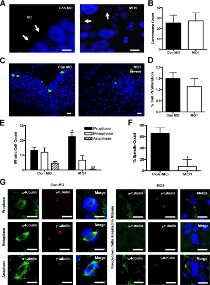
RASSF7 is required for spindle formation and completion of mitosis in the neural tube. (A) Centrosome number is not affected by RASSF7 knockdown. γ-tubulin staining (red) was used to examine the centrosomes (highlighted by arrows) of the neural tube of Con MO– and MO1-injected embryos and were counterstained with DAPI (blue). VC, ventricular cavity. (B) Centrosome number in the neural tube is unaffected by RASSF7 knockdown. No significant difference was found between Con MO and MO1; n = 6 specimens for each MO from three independent experiments. (C) Cellular proliferation was examined in the neural tube of MO-injected embryos by staining with anti-phospho-S10 histone H3 (green) and DAPI (blue). (D) The number of phospho-histone H3 proliferating cells as a percentage of the total number of cells counted (DAPI stain). Proliferation is similar between the two MO tissues and is not significantly different. n = 9 from three injection experiments for both MOs, >7000 cells were counted for each individual specimen. (E) Counts of dividing cells in the following phases: prophase (also includes prometaphase), * p< 0.05; metaphase, no significant difference; and anaphase (also includes telophase), ** p< 0.01. Mitotic phases were distinguished by DAPI staining using the following criteria: prophase/prometaphase, condensed DNA; metaphase, genetic material aligned on the metaphase plate; and anaphase/telophase, chromosome separation. Counts were from three experiments (Con MO, n = 90; MO1, n = 90). All error bars, SD. Knockdown cells arrest in early mitosis. (F) The total number of spindles counted as a percentage of the total number of dividing cells. * p< 0.05, n = 6 specimens for each MO, from three experiments. The number of mitotic spindles was greatly reduced in the dividing RASSF7 knockdown cells. (G) α-tubulin staining (green) was used to visualize spindles in mitotic cells of the neural tube, whereas γ-tubulin staining was used to mark the centrosomes (red). Sections were counterstained with DAPI (blue). Mitotic spindles were missing, or occasionally abnormal, in the dividing RASSF7 knockdown cells. All bars, 10 μm. RASSF7 knockdown cells fail to progress through mitosis because of a deficiency in spindle formation.

