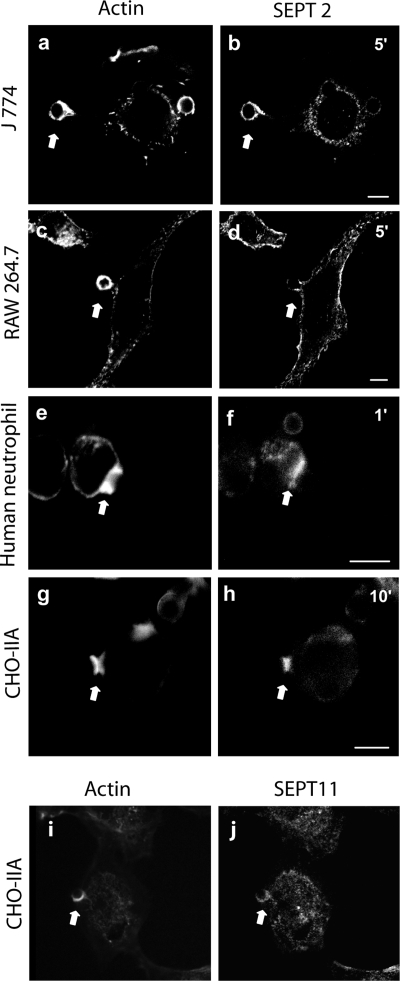
Figure 2.
Endogenous SEPT2 and SEPT11 accumulate on phagosomes in different phagocytic cells. Opsonized beads were allowed to attach to the cells for 10 min on ice and then RAW 264.7 (a and b), J 774 (c and d), human neutrophil (e and f), and CHO-IIA cells (g–j) were warmed to 37°C to start phagocytosis for 5, 5, 1, and 10 min, respectively, at 37°C. Cells were then fixed and endogenous SEPT2 was stained with rabbit polyclonal anti-human SEPT2 antibody (b, d, f, and h) or SEPT11 with anti-SEPT11 (j) and detected with Cy3-conjugated donkey anti-rabbit antibody. Actin was stained with Alexa 488-phalloidin (a, c, e, g, and i). The arrows indicate accumulated actin and septin at sites of interaction with opsonized beads in each cell line. Bars, 5 μm.

