
Figure 5.
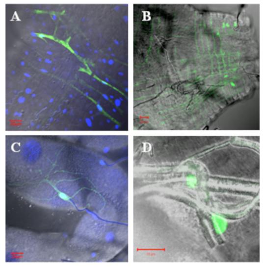
Confocal micrographs of dissected tissues from Ae. taeniorhynchus mosquitoes IT infected with GFP-expressing VEEV strain 3908 (A and B) and strain 68U201 (C and D) replicon particles. Transmitted image is overlayed with GFP fluorescent image. DAPI staining was added for images A and C. A) Circular muscles of the anterior midgut expressed GFP. B) Circular and longitudinal muscles of the posterior midgut expressed GFP. C) Tracheoles associated with the Malpighian tubules expressed GFP. D) Cells associated with the tracheoles of the midgut expressed GFP. Red bar in lower corners = 20μm.