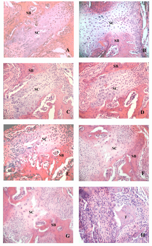
Figure 2.

Photomicrographs of midpalatal sutures during ME. A: 0 d control group, B: 9 d control group, C: 0 d ME group, D: 1 d ME group, E: 3 d ME group, F: 5 d ME group, G: 7 d ME group, and H: 9 d ME group. SC: suture cartilage. SB: suture bone. There were no significant differences between the two groups in the tissue structure of the midpalatal sutures before day 5, but from day 5 until the end of the study, there was significant tissue remodeling observed in the sutures of the ME group that was not observed for the control animals. (Haematoxylin-eosin, original magnification ×40). The tissue marked " T " was further validation as bone or cartilage by determination of Collagen I localization (Figure 5).
