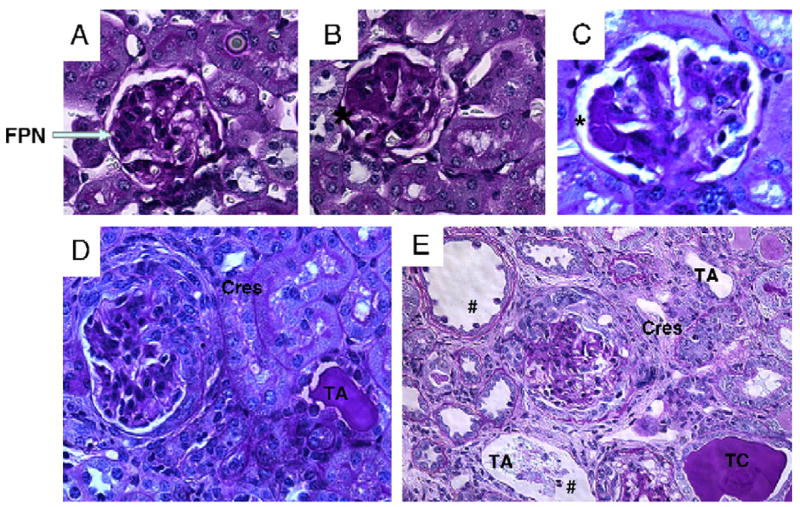
Figure 2.

Representative renal histology from pristane-induced lupus SJL/J mice. Mice were injected with pristane at 6 weeks of age and sacrificed at 32 weeks. Kidneys were harvested, fixed in 4% formalin and paraffin sections were stained with H&E and PAS. (A, D) Representative sections from male and (B, C, E) female mice are shown. These figures show typical epithelial and endothelial deposits (denoted by *), focal proliferative nephritis (FPN), diffuse glomerular hypercellularity (in D and E), cellular and fibrous crescents (Cres), tubular dilatation and atrophy (TA) and casts (TC) and tubular epithelial cell and macrophages (#) in tubular lumen. Summary of disease grades with differences in female versus male is shown in Fig. 3.
