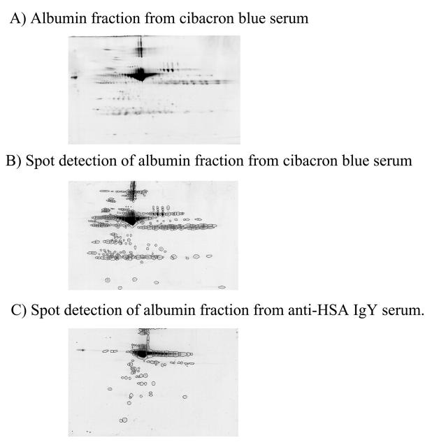
FIGURE 4.
Comparison of specificity of anti-HSA IgY antibodies and Cibacron blue for rat serum albumin. Seventy-five micrograms of Cibacron blue (albumin)-associated protein was subjected to 2DE on a pH 3–10 immobilized pH gradient strip and a 6%–15% SDS-containing polyacrylamide gel (A). The total number of spots in the albumin-associated fraction from both Cibacron blue (B, 304 spots) and anti-HSA IgY (C, 98 spots) were determined using Progenesis Discovery spot detection.

