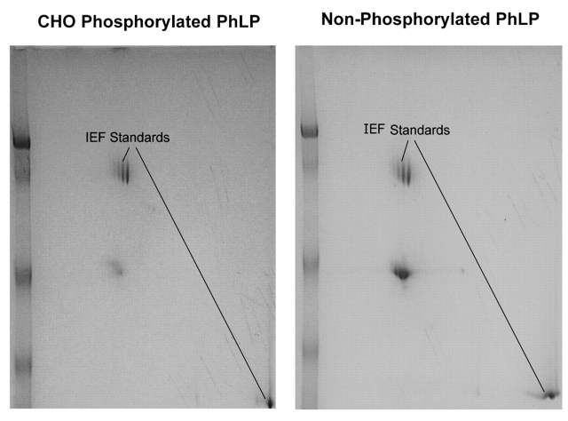
FIGURE 2.
2D Gel analysis of the stoichiometry of phosphorylation. Unphosphorylated and phosphorylated samples of phosducin-like protein were run on 2DE. Immobilized, 7-cm, pH 4–7 gradient strips were run on the Multiphor II apparatus for the first dimension. The second dimension was run on a 10% SDS-PAGE gel. The gel was stained with colloidal Coomassie. Gel images were captured using the AlphaDigiDoc system and analyzed using Melanie 4 software.

