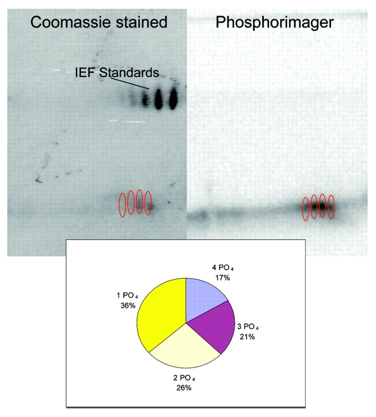
FIGURE 3.

2D Gel analysis of the stoichiometry of phosphorylation using 32P-γATP. The protocol was followed for phosducin-like protein phosphorylation with 32P-γATP. A 2DE gel was run and stained as in Figure 2, dried onto Whatman filter paper, and the radioactivity visualized using a phosphorimager. Melanie software was used to determine intensities of spots for phosphorylation comparison.
