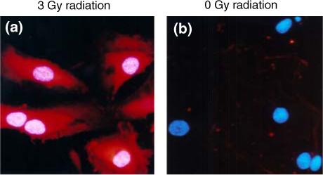
FIGURE 3.

Immunofluorescent microscopy of antibody binding to P-selectin in HUVECs treated with radiation. Cells were stained with DAPI nuclear stain (blue) and scFv 10A was conjugated with Cy3 dye (red). (a) scFv 10A binding to P-selectin on HUVECs treated with a radiation dose of 3 Gy. (b) No scFv 10A binding in HUVECs treated with a sham radiation dose of 0 Gy.
