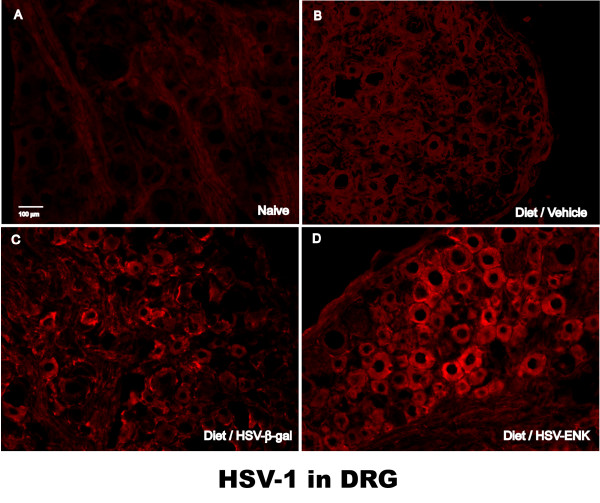
Figure 6.
HSV-1 immunohistochemical staining in thoracic DRG. Photomicrographs of immunohistochemical staining for human HSV-1 protein at week 10. No stain is evident in dorsal root ganglia of (A) naïve animals or (B) vehicle-treated animals fed the alcohol and high-fat diet. Note the presence of staining for human HSV-1 protein in DRG of animals given the (C) HSV-β-gal and (D) HSV-ENK vector treatments. Cytoplasmic localization is noted for HSV in DRG from vector treated animals only in lower thoracic ganglia (T7–T9).

