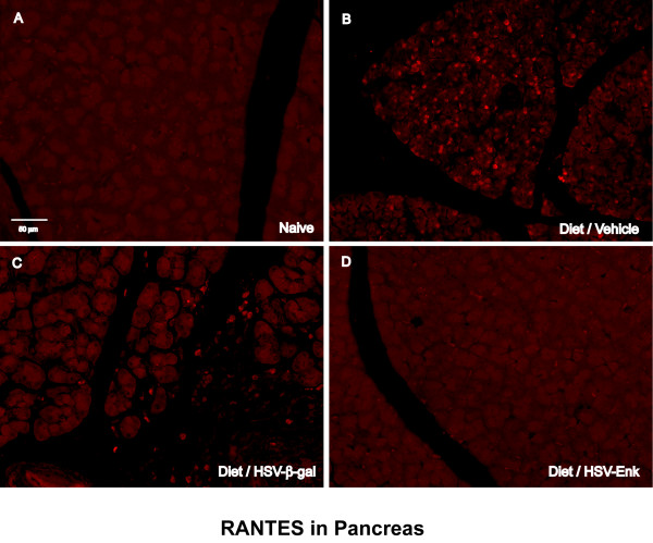
Figure 9.
RANTES immunohistochemical staining in pancreas. Photomicrographs of immunohistochemical staining of RANTES in the pancreas of rats are shown for week 10. A. Naïve rat pancreas. B. Diet-induced pancreatitis and application of vehicle as control. C. Diet-induced pancreatitis and application of the HSV-β-gal control vector. D. Animals given the same diet and application of HSV-ENK. Note the increased RANTES staining in pancreata of animals with alcohol and high-fat diet induced pancreatitis treated with vehicle or HSV-β-gal applications. Little or no staining of RANTES is noted in naïve and HSV-ENK-treated animals.

