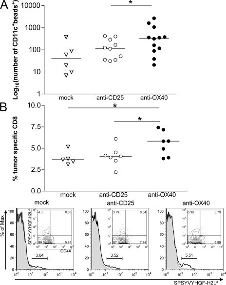
Figure 8.
T reg cell inhibition by OX40 triggering allows TIDC maturation and priming of new CTLs. (A) Rescue of DC migration after T reg cell inhibition. FITC-conjugated latex particles (2 × 10⁷ beads) of 1 μm in diameter were coinjected with anti-OX40, anti-CD25, or rat IgG (mock) within tumors of 3–4 mm in diameter. After 24 h, axillary and inguinal draining lymph nodes were collected and treated with collagenase D, and the obtained cell suspensions were stained for CD11c. The number of CD11c+ cells showing green fluorescence because of microbead engulfment was evaluated by flow cytometry. Each symbol corresponds to a single draining lymph node; the solid line represents the median value. Results are a pooled representation of three independent experiments. *, P < 0.05. (B) Induction of tumor-specific CD8+ T lymphocytes. (top) Tumor-bearing mice received intratumor injection of anti-CD25, anti-OX40, or rat IgG (mock). 5 d after treatment, axillary and inguinal draining lymph nodes were collected and stained for flow cytometry. On gated CD8+ T cells, the percentage of tumor-specific clones was evaluated by staining with H2Ld tetramers recognizing the SPSYVYHQF peptide of the gp70-env tumor-associated antigen. As a background staining, H2Ld tetramers specific for the unrelated peptide TPHPARIGL (E. coli β-galactosidase 876–884) were used. Percentages of tumor-specific CD8+ T cells were calculated by subtracting the background (β-galactosidase) from the specific (gp70-env) staining. OX86-treated mice showed the highest percentage of tumor-specific CD8+ T cells in the draining lymph nodes. Each symbol represents the draining lymph node of randomly chosen mice from two independent experiments. The solid line represents the median value. *, P < 0.05. (bottom) Representative histograms of tetramer-stained cells gated on CD8+ T cells are shown (indicated as percentages). Gp70-H2Ld tetramer staining (continuous line) was overlaid with β-galactosidase–H2Ld control tetramer staining (shaded line). The subtracted value is indicated for each sample. (insets) Surface expression of the T cell memory marker CD44 on gp70-H2Ld tetramer–positive CD8+ T cells.

