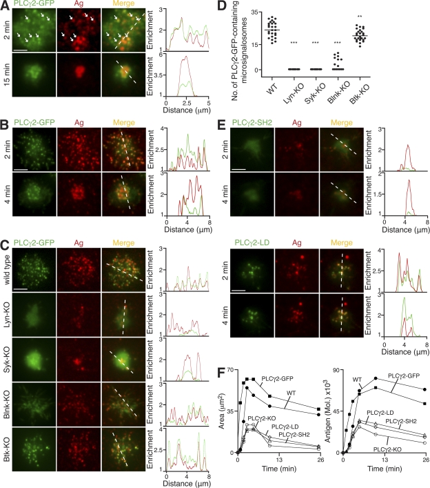
Figure 4.
Molecular requirements for PLCγ2 recruitment to antigen microclusters. (A–F) B cells were settled onto bilayers containing anti-κ (A) and anti-IgM (B–F) as antigen (Ag; red). (A–C and E) TIRFM images. Relative fluorescence intensity plots to indicate the distribution of antigen and various PLCγ2-GFP proteins (green) are depicted by the diagonal dashed lines. (A) Primary B cells from PLCγ2-GFP knock-in mice. White arrows identify selected antigen microclusters containing PLCγ2-GFP. (B) PLCγ2-KO DT40 B cells stably expressing PLCγ2-GFP. (C) WT, Lyn-KO, Syk-KO, Blnk-KO, and Btk-KO DT40 B cells expressing PLCγ2-GFP (green) after 3 min. (D) Quantification of PLCγ2-GFP–containing microsignalosomes present in WT, Lyn-KO, Syk-KO, Blnk-KO, and Btk-KO DT40 B cells after 3 min. Data are representative of two experiments, and the number of antigen microclusters containing PLCγ2-GFP was measured in 15–27 cells of each cell type, representing a total of 1,344 microclusters. Mean numbers are as follows: WT, 25 ± 1; Lyn-KO, 0; Syk-KO, 0; Blnk-KO, 3 ± 1; and Btk-KO, 21 ± 1. **, P < 0.005; ***, P < 0.0001. (E) PLCγ2-KO DT40 B cells stably expressing (top) PLCγ2-GFP with mutated SH2 domains (PLCγ2-SH2) or (bottom) PLCγ2-LD-GFP (PLCγ2-LD). (F) Quantification of (left) contact area by confocal microscopy and (right) antigen accumulation. Bars: (A) 2.5 μm; (B, C, and E) 5 μm. Videos 5–7 are available at http://www.jem.org/cgi/content/full/jem.20072619/DC1.

