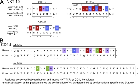
Figure 3.
Sequence alignment of CD1d and the NKT TCR from both human and mouse. Sequence comparison of the CDR1α, CDR3α, and CDR2β loops of the mouse and human NKT TCR homologues (A) and the CD1d α1- and α2-helices (B). Substituted residues that have a >10-fold reduction in affinity are shown in red; a 4–6-fold reduction in affinity are shown in green; and a <4-fold reduction in affinity are shown in blue; residues that contact either CD1d–α-GalCer or the NKT TCR that were not substituted, purple.

