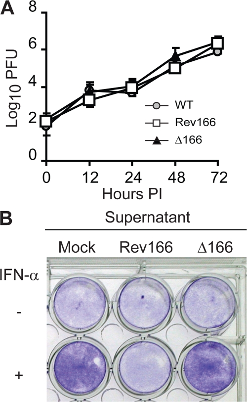
Figure 1.
In vitro characterization of Δ166 and Rev166 ECTV. (A) Mouse fibrosarcoma A9 cells were infected with 0.01 PFU/cell of the indicated viruses. Cell-associated and free virus were determined at the indicated times after infection. Data points are means ± SD of three replicate wells. (B) Confluent L929 cells in 24-well plates were treated with 200 μl of supernatants from BSC-1 cells infected as indicated. After 1 h of incubation, 300 μl of media with or without 50 IU IFN-α was added. The cells were incubated for an additional 24 h, followed by infection with 10 PFU/cell VSV. After 24 h of incubation, the cells were washed, fixed, and stained with crystal violet.

