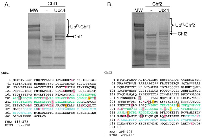
Figure 5.
Identification of Ubc4 and Ubc13/Mms2-dependent Chf protein sites of autoubiquitination. Coomassie-stained gels of purified Ubc4-dependent autoubiquitination reactions are shown above the sequences of Chf1 (A) and Chf2 (B). As described in Materials and Methods and as shown in Table 2, sites of ubiquitination were identified by tandem mass spectrometry. Lys residues are shown in magenta, with sites ubiquitinated by Ubc4 in gray and sites ubiquitinated by both Ubc4 and Ubc13/Mms2 in yellow. Sites ubiquitinated in trans reactions are boxed. FHA domains are in green and RING domains are in blue.

