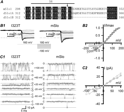
FIGURE 1.
The kinetic characteristics of both the instantaneous and single-channel currents in the mSlo and mutant I323T. (A) Sequence alignment within the pore region of mSlo, dSlo (A2, C2, E2, G5, 10) splice variant (dSlo1A) (1,23) and dSlo1B (NM_001014653). The horizontal line indicates the S6 segment predicted as the pore region. The next 20 amino acids after S6 form the linker in BK-type channels. Identical residues are boxed in dark gray, and similar amino acid residues in light gray. The arrowhead in the S6 domain indicates the mutated amino acids used in this study. (B1) Traces show instantaneous currents obtained from the inside-out patches from Xenopus laevis oocytes injected with cRNA encoding the mSlo (right, n = 5) or its mutant I323T (left, n = 7), separately. Currents were activated by a voltage step to +160 mV, followed by steps to potentials from −180 mV to +160 mV in 10-mV increments, in symmetrical 160 mM K+ solutions with 10 μM intracellular Ca2+. The voltage protocol is shown at the bottom middle. Traces in thick black lines represent currents at +100 and −100 mV, respectively. (B2) The normalized amplitudes of instantaneous currents (± SD) are plotted as a function of voltages for mSlo (▿) and I323T (○). (C1) The representative single-channel currents of mSlo and I323T were obtained at +100, +60, +20, 0, −20, −60, and −100 mV, respectively. All the single-channel recordings were digitized at 20 kHz and filtered at 10 kHz. Solid lines labeled with “c” or “o” represent the closed and open levels, respectively. (C2) The amplitudes of single-channel currents for mSlo (▿) and I323T (○) are plotted as functions of holding potentials.

