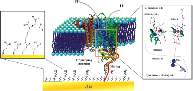
FIGURE 1.
Schematic representation of CcO embedded into a protein-tethered bilayer lipid membrane. The protein is attached to the surface of a template stripped gold film by the his-tag attached to SU II, allowing for direct ET. The lipid bilayer is assembled around the protein. The box on the right shows the stepwise ET inside the CcO (44) and the box on the left shows the details of the surface modification (11).

