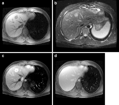
Fig. 2.
A 14-year-old boy after resection of a bronchial NEC. Axial T1-W image (a) demonstrates a hypointense liver lesion that is moderately hyperintense on the T2-W image (b). Arterial phase image after gadolinium administration (c) shows intense early enhancement with wash-out to isointensity on the portal venous phase images (d), consistent with hypervascular metastasis

