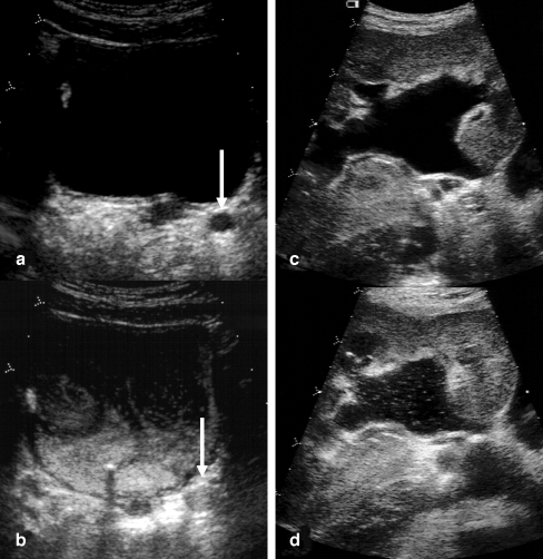
Fig. 1.
Scans in fundamental mode before (a, c) and after (b, d) contrast agent administration of a dilated left distal ureter (a, barrow) and pelvicalyceal system (c, d). In the postcontrast scans echogenic microbubbles fill the distal ureter (b) and are also detected in the pelvicalyceal system (d)

