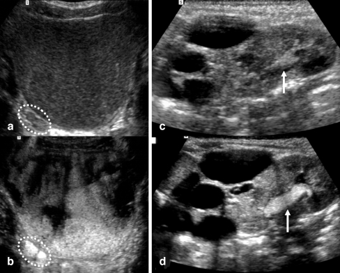
Fig. 2.
Scans after contrast agent administration in fundamental mode (a, c) and with harmonic imaging (b, d) of the bladder and right dilated ureter (a, bdotted circle) and a duplex kidney (c, d) with a multicystic dysplastic upper moiety. Reflux in the right ureter and in the lower moiety of the duplex kidney (grade II, arrow) are much more conspicuous with harmonic imaging (b, d). Note also the crisper depiction of the cysts in the upper moiety with harmonic imaging

