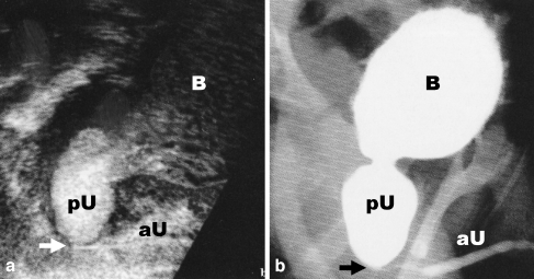
Fig. 5.
Transperineal voiding urethrosonography (a) as part of VUS in comparison with (b) VCUG. To facilitate the comparison the US image (a) is presented upside down. Note in the transperineal US (a) the microbubbles in the bladder (B) and in the massively dilated posterior urethra (pU). The anterior urethra (aU) is depicted as very thin in the presence of a posterior urethral valve (arrow). The finding was confirmed on VCUG (b) (courtesy of Dr. M. Bosio, Milan, Italy)

