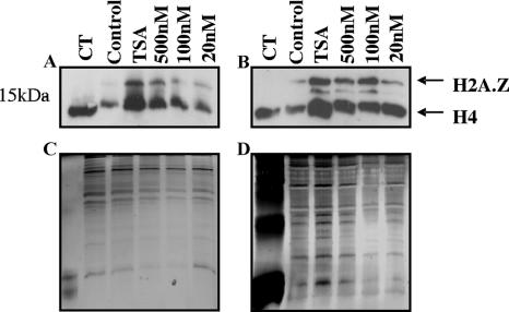
FIG. 4.
Hyperacetylation of P. falciparum histones. P. falciparum-infected erythrocytes (Dd2) were cultured in vitro in the presence of TSA (500 nM) or 500 nM, 100 nM, and 20 nM of compound 9 (A and C) or compound 14 (B and D) for 3.5 h. Matched controls received no drug (control) (0.1% DMSO). Histones were acid extracted in 0.5 M HCl and separated on 15% SDS-polyacrylamide gels. Hyperacetylation was determined by Western blotting using polyclonal anti-tetra-acetyl histone H4 antisera (Upstate) (A and B). Silver staining was carried out as a loading control (C and D). Arrows indicate different histones. CT, calf thymus histones.

