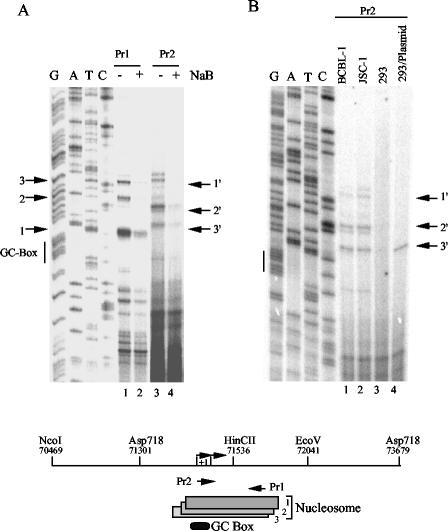
FIG. 5.
Primer extension mapping of Mnase I nuclease cleavage sites in ORF50. (A) Mononucleosomal fragments from Mnase-treated nuclei were gel purified and analyzed by primer extension with primers Pr1 and Pr2, as indicated above the gel. Sequence reactions were generated with Pr1, and the GC box is indicated to the left. Numbered arrows indicate the major primer extension products that correlate with Mnase I cleavage sites and nucleosome boundaries. (B) Primer extension with Pr2 was used to compare the mononucleosomal fragments derived from BCBL-1, JSC-1, and 293 cells and 293 cells transfected with ORF50-Luc plasmid DNA. Sequencing lanes were generated with Pr1. The schematic at the bottom indicates the nucleosome positions mapped by the above primer extension and analysis.

