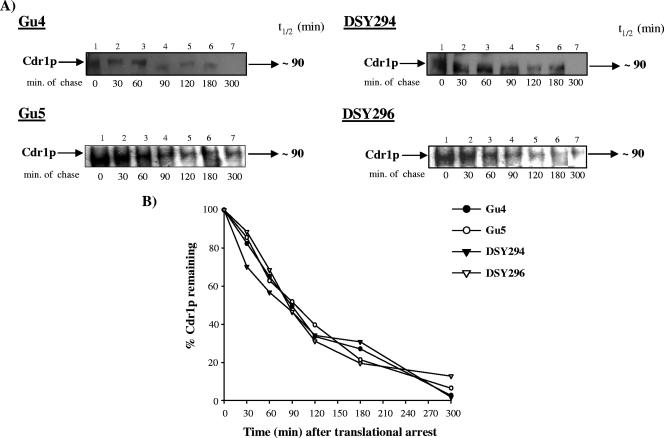
FIG. 7.
Cdr1p decay assay. (A) Exponentially grown cultures of C. albicans were translationally halted at 30°C by addition of 75 mM of cycloheximide for 1 h. Whole-cell extracts were prepared at the indicated times after cycloheximide treatment. For AR isolates, ∼20 μg, and for AS isolates, ∼30 μg (because of relatively low expression of Cdr1p) of crude extract for each time was loaded and separated by SDS-polyacrylamide gel electrophoresis. Equal loading of protein was assessed using a Coomassie-stained gel (data not shown). Cdr1p was detected using a polyclonal anti-Cdr1p antibody. The Cdr1p-specific bands were subsequently quantified by densitometry scanning in a phosphorimager. (B) Band intensities (represented as percentages of the value at T0) for each isolate were plotted against the chased time. t1/2, half-life.

