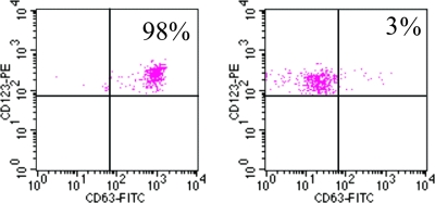
FIG. 5.
Human BAT in allergic and normal control subjects. Activation of basophils was detected by flow cytometry using a fluorophore-tagged (phycoerythrin [PE]) MAb against the CD63 cell activation marker. The expression of CD63 (horizontal axis) is plotted against levels of anti-IgE fluorescein isothiocyanate (FITC) (vertical axis) in response to A. simplex crude extract. The left-hand plot shows results from the first patient described 11 years before (25), although the test was carried out 11 years later (2006), and the right-hand panel corresponds to a female subject exhibiting no allergy to A. simplex used as a control. The results are expressed as the percentage of CD63+ basophils. Note that activated basophils from the allergic patient appear in the upper right window encompassing 98% of marked cells. In contrast, the control (right-hand plot) shows that a mere 3% of basophils were activated upon exposure to parasite antigen/allergens. (M. T. Audicana and N. Longo, unpublished data.)

