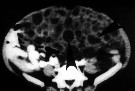
FIG. 16.
CT scan, demonstrating vesicles of Echinococcus vogeli in the abdomen (from the 58-year-old female case from reference 12). The vesicles are hypodense and round to oval. The intestine has been displaced against the posterior wall. (Reprinted from reference 12 with permission from Elsevier.)

