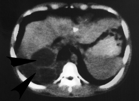
FIG. 19.
CT scan, made 5 years after surgery (from the 22-year-old male case from reference 12), showing hypodense, round polycystic vesicles, mainly posterior in the right hepatic lobe and right lung. Triangular calcifications are evident in the liver. (Reprinted from reference 12 with permission from Elsevier.)

