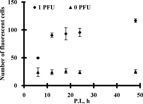
FIG. 3.
The numbers of fluorescent cells at different p.i. time points. FRhK-4 cells infected with 0 or 1 PFU for 6, 12, 18, 24, and 48 h p.i. were fixed and permeabilized before 1 h of incubation with 5 μM MB H1. The number of fluorescent cells was counted using a fluorescence microscope at ×100 magnification. Error bars represent the standard deviations for three replicates.

